Dorsal Root Entry Zone Lesioning means that the neurosurgeon makes a lesion within the dorsal root entry zone, which is the first important level of modulation for pain sensation. DREZ lesioning is a last effort technique for patients suffering from refractory chronic pain due to brachial plexus injuries (especially those with avulsion), spinal cord lesions (predominantly in the conus medullaris), segmental pain caused by lesions in the cauda equina, peripheral nerve injuries, amputation, or herpes zoster. Lesioning techniques include microsurgical coagulation, radiofrequency thermocoagulation, laser beam or ultrasound lesion maker.
In our institute we mainly focus on pain after brachial plexus avulsion (BPA) which is generally characterized by 2 main different components: paroxysmal (electrical shooting-like) pain, and continuous background (burning) pain. The technique is more effective on the paroxysmal component than on the continuous component of pain.
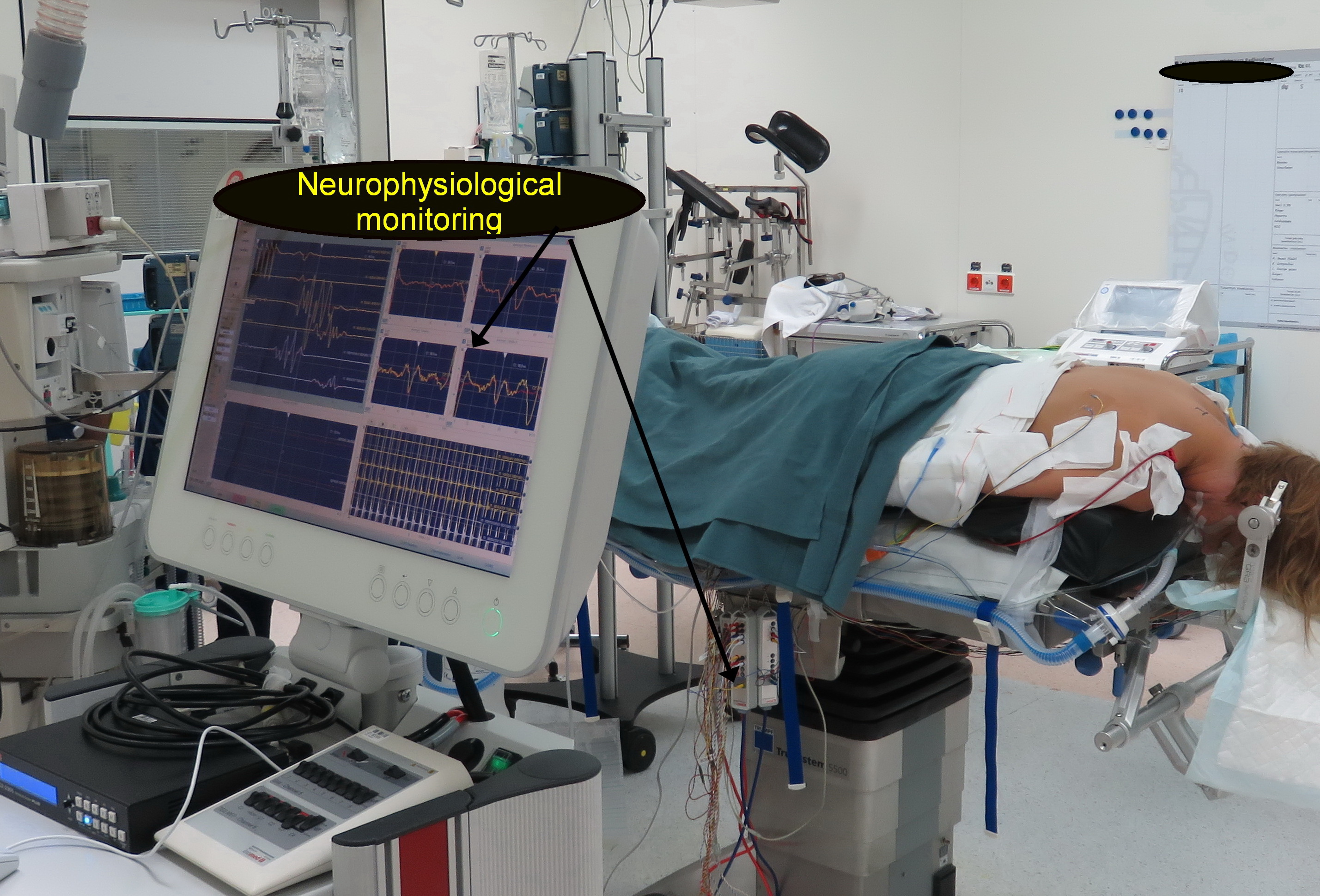
The procedure implies cervical (hemi-) laminectomy (general anasthesia, prone position, mayfieldclamp, intraoperative X ray, micrrroscope). During surgery intraoperative neuromonitoring is performed using SSEP, MEP and dorsal column stimulation to localize the midline and the dorsal root sulcus which might be very difficult to recognize due to the avulsion of the roots that leads to abnormal anatomy of the spinal cord (distortion, rotation, hemi atrofia, formation of meningocele). Under microscopic magnification a longitudinal incision is made in the dorsolateral sulcus ventrolaterally at the entrance of the rootlets into the sulcus. Microbipolar coagulations or lesions are performed continuously inside the sulcus down to the apex of the dorsal horn (Rexed’s layers I-V) and medial part of the tract of Lissauer (TL), where the afferent fibers synapse with the cells of the sensory spinoreticulothalamic ascending pathways. The region can be recognized by its brown-gray colour. The average lesion is 2 to 3 mm deep and is made at a 35° angle medially and ventrally.
The procedure is presumed to preferentially destroy the nociceptive fibers grouped in the lateral bundle of the dorsal rootlets, as well as the excitatory medial part of the Tract of Lissauer. The upper layers of the dorsal horn are also destroyed if coagulations or lesions are made inside the dorsal horn . The upper layers of the dorsal horn are known to be the site of “hyperactive” neurons, especially in the cases with peripheral deafferentation.
The procedure is presumed to at least partially preserve the inhibitory structures of the DREZ, (i.e., the lemniscal fibers reaching the dorsal column, as well as their recurrent collaterals to the dorsal horn and the substantia gelatinosa [SG] propriospinal interconnecting fibers running through the lateral part of the TL).